

Introduction

Degrader Antibody Conjugates (DACs) represent a revolutionary therapeutic modality that combines the precision of monoclonal antibodies (mAbs) with the potent protein degradation capabilities of targeted protein degraders, such as proteolysis-targeting chimeras (PROTACs) or molecular glues. This hybrid approach leverages the strengths of both technologies: the ability of antibodies to selectively deliver payloads to specific cells or tissues, and the capacity of degraders to induce the ubiquitination and subsequent proteasomal degradation of disease-causing proteins. DACs hold immense promise for addressing previously “undruggable” targets, particularly in oncology, autoimmune diseases, and neurodegenerative disorders (1-15). However, their development is accompanied by significant scientific and technical challenges. This essay provides a comprehensive exploration of the opportunities, innovations, and challenges associated with DACs, integrating insights from recent scientific advancements and studies.
What are Degrader Antibody Conjugates (DACs)
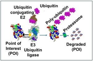
Degrader Antibody Conjugates (DACs) are an emerging class of targeted therapeutics that combine the specificity of monoclonal antibodies with small-molecule targeted protein degrader (TPD), such as proteolysis-targeting chimeras (PROTACs) or molecular glues. DACs comprise three key components. The targeting antibody binds with high specificity to cell surface antigens such as HER2 or CD33, ensuring precise delivery. The chemical linker is often cleavable, using mechanisms like pH sensitivity or protease cleavage, allowing the controlled intracellular release of degraders. Lastly, the degrader moiety, typically PROTACs or molecular glues, recruits E3 ligases such as cereblon or VHL to facilitate ubiquitination and subsequent protein degradation. This catalytic mechanism allows DACs to degrade traditionally “undruggable” proteins, such as transcription factors or scaffold proteins, with high specificity and reduced off-target effects . DACs hold promise not only in oncology, where they can degrade oncogenic proteins like HER2 or KRAS mutants, but also in neurodegenerative diseases, autoimmune disorders, and beyond.
Comparison of Degrader Antibody Conjugates (DACs), Targeted Protein Degraders (TPDs), like PROTACs, and Antibody-Drug Conjugates (ADCs)
Targeted therapeutics have advanced with Degrader Antibody Conjugates (DACs), Targeted Protein Degraders (TPDs) like PROTACs, and Antibody-Drug Conjugates (ADCs). These approaches differ in design, function, and application. DACs merge antibody specificity with protein degradation. TPDs, such as PROTACs, use bifunctional molecules to recruit E3 ligases for proteasomal degradation. ADCs link antibodies to cytotoxic drugs for targeted delivery.
A detailed comparison of degrader antibody conjugates (DACS), PROTACs, and antibody-drug conjugates (ADCs), are shown in the following Table (Table 1). This comparison examines their mechanisms, benefits, and challenges, aiding researchers in selecting the best approach for precision medicine and improved patient outcomes.
Table 1: Comparison of DACSs, PROTACs, and ADCs
| Feature | Degrader Antibody Conjugates (DACs) | PROTACs / Molecular Glues | Antibody Drug Conjugates (ADCs) |
|---|---|---|---|
| Targeting Mechanism | Antibody-mediated binding to specific proteins or cell surface receptors | Small-molecule-mediated recruitment of E3 ubiquitin ligase to target proteins | Antibody-mediated binding to specific cell surface antigens |
| Structure | Antibody linked to a degrader (e.g., PROTAC) via a chemical linker. | Bifunctional molecule: one end binds the target protein, the other recruits an E3 ubiquitin ligase. | Antibody linked to a cytotoxic drug via a chemical linker. |
| Delivery Method | Delivered via monoclonal antibody conjugation | Diffuses across cell membranes | Delivered via monoclonal antibody conjugation |
| Target Selectivity | Highly specific due to antibody targeting | Depends on small-molecule selectivity and cellular permeability | Highly specific due to antibody targeting |
| Mechanism of Action | Antibody binds to target, internalizes or releases degrader, leading to ubiquitination and proteasomal degradation | Directly brings target protein and E3 ligase into proximity for ubiquitination and degradation | Antibody binds to target, internalizes, and releases cytotoxic payload to kill the target cell |
| Catalytic Activity | A single degrader can induce degradation of multiple target protein molecules. | Operates catalytically to degrade multiple target protein molecules. | Each payload molecule acts stoichiometrically (one payload molecule kills one cell). |
| Target Proteins | Can target both extracellular and intracellular proteins (via antibody-mediated delivery). | Primarily targets intracellular proteins. | Primarily targets extracellular proteins or cell surface receptors. |
| Undruggable Targets | Can degrade proteins lacking enzymatic activity or binding pockets (e.g., transcription factors). | Limited to proteins with accessible extracellular domains or cell surface receptors. | Can degrade proteins lacking enzymatic activity or binding pockets (e.g., transcription factors). |
| Specificity | High specificity due to antibody-antigen binding; reduces off-target effects. | Moderate specificity; depends on the binding affinity of the degrader for the target protein. | High specificity due to antibody-antigen binding; reduces off-target effects. |
| Internalization Requirement | May require internalization for action on intracellular targets | No internalization required, acts intracellularly | Internalization is required for cytotoxic payload release |
| Degrader/Payload Release | Degrader is cleaved or released inside the cell or extracellularly | Degrader acts freely upon entering the cell | Cytotoxic payload is cleaved and released inside the cell |
| Therapeutic Applications | Oncology, neurodegenerative diseases, autoimmune disorders, infectious diseases. | Primarily oncology (e.g., HER2-positive breast cancer, CD30-positive lymphomas). | Oncology, neurodegenerative diseases, autoimmune disorders. |
| Systemic Exposure | Localized delivery reduces off-target toxicity | Small molecules can have broader distribution and potential off-target effects | Localized delivery reduces off-target toxicity but depends on linker stability |
How to design Mechanism of Action (MOA) assays for degrader antibody conjugates (DACs)
Designing Mechanism of Action (MOA) assays for degrader antibody conjugates (DACs) involves understanding their unique structure, which combines the specificity of antibodies with the protein degradation capabilities of degraders like PROTACs or molecular glues. DACs consist of three components: an antibody targeting a cell surface antigen, a linker, and a degrader moiety. MOA assays must evaluate each component’s role in the DAC’s function.
Key steps in designing MOA assays include:
1. Target Binding: Assessed using flow cytometry, SPR, or ELISA to confirm antigen interaction.
2. Internalization: Tracked via confocal microscopy, pH-sensitive dyes, or flow cytometry.
3. Degrader Release: Monitored using mass spectrometry, fluorogenic assays, or Western blotting.
4. Target Protein Engagement: Evaluated with CETSA, PLA, or Co-IP to confirm interaction with the ubiquitin-proteasome system.
5. Protein Degradation: Quantified using Western blotting, qPCR, or ubiquitination assays.
6. Functional Consequences: Measured through assays like CellTiter-Glo, phospho-specific antibody analysis, or apoptosis assays.
Specificity and minimizing off-target effects are critical. Control experiments, CRISPR/Cas9 knockout validation, and proteomics-based analysis help confirm target dependency and detect unintended degradation. Assay optimization involves dose-response curves, time-course experiments, and selecting appropriate cell lines.
Pharmacodynamic (PD) biomarkers should link target degradation to functional outcomes, aiding the transition from in vitro to in vivo studies. In vivo evaluation includes xenograft models, pharmacodynamic assays, and imaging techniques to assess efficacy, protein degradation, and biodistribution.
Key considerations include linker stability, degrader potency, and antibody specificity. By systematically addressing each mechanistic step, robust assays can be developed to evaluate DAC efficacy, specificity, and overall MOA.How to design in vitro and cell-based potency assays for degrader antibody conjugates (DACs).
How to design in vitro and cell-based potency assays for degrader antibody conjugates (DACs)
Designing potency assays for degrader antibody conjugates (DACs) requires a deep understanding of their mechanism of action (MOA) and the ability to quantitatively measure their biological activity. DACs combine the precision of antibodies with the protein degradation capabilities of degraders like PROTACs or molecular glues, making their potency assays more intricate than those for traditional antibodies or small molecules. These assays must evaluate critical parameters, including target binding, internalization efficiency, degrader release, target protein degradation, and functional outcomes. Key metrics such as DC50 (concentration for 50% degradation), IC50 (concentration for 50% inhibition of a functional readout), and Dmax (maximum degradation level) are used to quantify these parameters.
Selecting a suitable cell line is crucial, ensuring it expresses the target antigen and protein at levels relevant to the disease context. Techniques like flow cytometry and Western blotting help validate antigen expression and DAC sensitivity. The primary measure of DAC potency is target protein degradation, which can be assessed through Western blotting for quantification, quantitative immunofluorescence for single-cell visualization, flow cytometry for high-throughput analysis, and mass spectrometry for proteomics-based quantification and off-target detection.
Functional assays are used to evaluate the downstream biological effects of target protein degradation. Cell viability assays, such as MTT or CellTiter-Glo, determine IC50 values by measuring proliferation or survival. Apoptosis can be assessed using Annexin V staining, caspase activation, or TUNEL assays, while cell cycle analysis via flow cytometry identifies changes like G1 arrest. Pathway-specific assays, such as those using phospho-specific antibodies or reporter constructs, measure signaling changes downstream of the target protein.
To ensure assay accuracy, controls are essential, including negative controls (non-targeting antibodies or degrader-free conjugates) and positive controls (known degraders or inhibitors). CRISPR/Cas9 knockout experiments confirm target dependency. Optimization involves generating dose-response curves across a range of DAC concentrations (e.g., 0.1 nM to 10 µM) to determine potency metrics, conducting time-course experiments to assess degradation kinetics, and optimizing cell density for assay robustness.
Assay validation focuses on reproducibility, sensitivity, and specificity, ensuring results reflect DAC activity rather than nonspecific effects. Pharmacodynamic (PD) biomarkers are identified to correlate target degradation with functional outcomes, aiding the transition from in vitro to in vivo potency assessments. In vivo potency assays include xenograft models to measure tumor growth inhibition and target protein degradation, pharmacodynamic assays to track biomarker changes in tumor or tissue samples, and imaging studies using fluorescent or radiolabeled DACs to evaluate biodistribution and target engagement.
Data analysis is performed using tools like GraphPad Prism to generate dose-response curves and calculate potency metrics (DC50, IC50, Dmax). Integrating in vitro and in vivo data provides a comprehensive understanding of DAC potency, ensuring a systematic approach to assay development. Key considerations include optimizing linker stability to balance circulation stability and intracellular cleavage, enhancing degrader potency for effective target engagement, and confirming antibody specificity to minimize off-target effects.
A typical DAC potency assay workflow involves seeding target-expressing cells, treating them with varying DAC concentrations, harvesting samples for Western blot or immunofluorescence, performing functional assays, and analyzing data to generate potency curves. By systematically addressing each step of the DAC MOA and incorporating rigorous controls and validation measures, robust potency assays can be designed to evaluate the efficacy and specificity of degrader antibody conjugates.
Challenges and opportunities in degrader antibody conjugates (DACs)
DAC development faces hurdles like endosomal entrapment, limiting intracellular degrader delivery and requiring innovative strategies. Immune responses to conjugates necessitate humanized antibodies or stealth coatings. Resistance mechanisms, such as target downregulation or E3 ligase mutations, complicate efficacy. Manufacturing is complex, demanding precise conjugation and quality control, while balancing antibody-degrader pharmacokinetics remains critical for minimizing toxicity.
Combining DACs with immune checkpoint inhibitors or chemotherapy shows promise, particularly in targeting PD-L1 with anti-PD-1 antibodies. AI and machine learning optimize DAC design and identify novel E3 ligase-target pairs. Emerging technologies like LYTACs and AUTACs expand DAC applications to extracellular or aggregated proteins. Clinical trials targeting HER2, EGFR, and CD20 are underway, offering insights into DAC safety and efficacy. These advancements highlight DACs’ potential in precision medicine.
Conclusion
Degrader Antibody Conjugates (DACs) represent a transformative advancement in targeted protein degradation, offering new hope for the treatment of cancers, neurodegenerative disorders, and other complex diseases. By integrating the specificity of monoclonal antibodies with the catalytic efficiency of protein degraders, DACs provide a novel and highly selective therapeutic approach. However, challenges such as intracellular delivery, immunogenicity, and manufacturing complexity must be addressed to fully realize their clinical potential. With continued innovation in antibody engineering, linker design, and degrader optimization, DACs are poised to revolutionize precision medicine and establish a new paradigm in targeted therapeutics.
References
- Degrader-antibody conjugates.
- Degrader-Antibody Conjugates: Emerging New Modality.
- New guise ADCs couple antibodies to degraders.
- A novel ROR1-targeting antibody-PROTAC conjugate promotes BRD4 degradation for solid tumor treatment.
- The New Frontier: Merging Molecular Glue Degrader and Antibody–Drug Conjugate Modalities To Overcome Strategic Challenges.
- What influences the activity of Degrader-Antibody conjugates (DACs).
- Delivery of a BET protein degrader via a CEACAM6-targeted antibody–drug conjugate inhibits tumour growth in pancreatic cancer models.
- A novel ROR1-targeting antibody-PROTAC conjugate promotes BRD4 degradation for solid tumor treatment
- Antibody–Drug Conjugates: A Start of a New Era in Gynecological Cancers.
- New-generation advanced PROTACs as potential therapeutic agents in cancer therapy.
- Allosteric antibodies: a novel paradigm in drug discovery.
- Single-Domain Antibody-Based Protein Degrader for Synucleinopathies.
- Drug conjugates for the treatment of lung cancer: from drug discovery to clinical practice.
- Diverse Roles of Antibodies in Antibody–Drug Conjugates.
- A novel ROR1-targeting antibody-PROTAC conjugate promotes BRD4 degradation for solid tumor treatment.
Our Recent Publication/Meeting Presentation
American Society of Hematology (ASH) Annual Meeting 2024.
Comprehensive Assay Solutions for In Vitro and Cell Based Potency Assays and Pharmacokinetics Studies- Our Expertise
With 30 years of expertise in cell culture, cell-based assays, and preclinical/clinical PK/PD analysis, we specialize in offering assay services essential for a wide variety of therapeutic drug development programs, preclinical studies, IND/BLA applications, and commercialization. Our comprehensive services include both preclinical non-GLP and GLP assays, as well as non-GMP and GMP assays, providing critical support throughout the entire development pipeline.
Watch the following video and explore our latest presentation on the development and validation of potency and pharmacokinetic (PK) assays for AAV vectors, highlighting innovative methodologies and industry-leading expertise.
Download the full presentation: Development of Custom Cell Based and In vitro Potency and Pharmacokinetics (PK) Assays for AAV vectors- Marin biologic Laboratories
Development of Cell-Based Potency Assays: Case Studies and Blogs from Marin Biologic Laboratories (MarinBio)




Drug Discovery & Development Assays Offered by Marin Biologic Laboratories (MarinBio)
- ADME/Tox
- Molecular Biology
- The Art of Cell Culture
- Exosomes
- Stability Services
- Potency Assay
- Gene Therapy Assays
- Immunotherapy Assays
- Antiviral Therapy Assays
- cGMP
- MLR
- Cell Based Assays
- ELISA
- Flow Cytometry
- Protein
- PCR-qPCR
- Immunoassay
- Radioimmunoassay
- GLP
- Transfection
- Cell Therapy Assays
- Targeted Protein Degradation
- Research to Commercialization
Our Recent Blogs/Articles
Design and Validate a GMP Cell Based Assay
Development and Validation of Cell-Based Potency Assays for AAV Therapies: Insights From a Commercial AAV Vector Study
Precision in Efficacy: Developing Robust GMP Potency Assays for PROTACs, Molecular Glues, and Other Targeted Protein Degradation (TPD) Drugs
PROTACs for Innovative Drug Development: Current Trends and Future Prospects
Advancing Bispecific Antibody Constructs: Potency Assay Innovations at MarinBio
Advancing PROTAC Therapeutics: Potency Assays for FDA Compliance
Cutting-Edge ELISA Techniques for Bispecific Antibodies by MarinBio
Advancing Cancer Therapeutics: The Role of Potency Assays in ADC Effectiveness
Radiopharmaceutical Drugs: Harnessing the Power of Targeted Radiation Therapy to Combat Cancer
Disclaimer: This information is intended solely for research purposes and does not serve as medical advice.
