

Contemporary and Mechanistically Distinct Novel Cancer Drugs
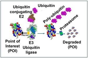
Recent advances in cancer therapeutics have ushered in a new era of precision oncology, with several novel drug classes demonstrating unique ways of killing cancer cells beyond traditional chemotherapies. The targeted protein degradation field has made significant strides, exemplified by the development of molecular glues (promotes target binding to E3 ligases and subsequent degradation) and bifunctional degraders (PROTACs) that induce the selective destruction of protein targets that standard therapeutics cannot inhibit. For instance, a novel RBM39 molecular glue has shown the ability to degrade the RBM39 protein, leading to complete tumor regression by impairing homologous recombination repair pathways.
Other innovative approaches include small molecule inhibitors that target specific metabolic pathways or protein-protein interactions, as well as antibody-drug conjugates (ADCs) that deliver cytotoxic agents directly to cancer cells, minimizing harm to healthy tissue. In addition there are drug modalities that affect intracellular signaling machinery including macrocyclic (circular) peptides and tumor suppressor stabilizers. These mechanistically novel therapies are rapidly moving toward clinical application, offering new hope for patients with cancers that have historically lacked effective targeted treatments. Progression of all cancer therapeutics into the clinic requires specific and robust in vitro cell based assays.
In Vitro Cell-Based Assays for Drug Development
Cell-based assays play a central role in modern drug development by enabling comprehensive evaluation of drugs across multiple stages of discovery and preclinical research. Cell-based assays assess potency and biological activity by measuring how effectively a drug exerts its intended effect in a cellular context. They are also essential for elucidating mechanisms of action, analyzing drug effects on cellular pathways, signaling, and gene expression, and for detecting off-target effects that could compromise drug safety.
Additional applications include monitoring cell proliferation and apoptosis (critical for cancer drug development), studying signal transduction and pathway modulation. Cell-based assays also facilitate the study of functional and biochemical endpoints, target engagement and saturation of the binding site, and resistance mechanisms, while supporting pharmacodynamic biomarker monitoring to optimize dosing regimens.
Overall, cell-based assays accelerate the drug development pipeline, enhance safety and efficacy evaluations, and provide biologically relevant data critical for bringing new therapeutics to market efficiently.
This report describes several mechanistically distinct non-immune modulating cancer therapeutics and the cell-based assays used to characterize them.
In Vitro Cell Based Assays for Cutting-Edge Cancer Therapeutics
Below is a list of cell-based assays used to move development of drugs across multiple stages of discovery and preclinical research. The assays listed meet requirements for each mechanistically distinct drug modality.
1. Cytotoxicity / Cell viability Assays
Cytotoxicity assays come in different formats and can apply to different mechanisms of cell 1) Detect toxic effects by measuring cell lysis, membrane integrity (e.g., LDH or Calcein release). 2) Measure the number of live cells in a sample using dyes (e.g., live: Calcein-AM; dead: Propidium iodide) or 3) metabolic indicators (e.g., resazurin, Celltiter Glo [CTG] measure of intracellular ATP).
- Antibody-drug conjugate. e.g. AbbVie ABBV-969
- Tumor suppressor stabilizer. e.g. Outrun Therapeutics OTX-2101
- Oncolytic virus. e.g. Replimmune OVX-101
2. Proliferation Assays
Proliferation assays can quantify cell division through a number of different techniques. Cell division in an in vitro assay is commonly measured using DNA content staining (e.g., propidium iodide or BrdU). Other assays include ATP-Based luminescence assays such as CellTiter-Glo, that quantifies ATP as a marker of metabolically active (viable) cells, generating a luminescent signal proportional to the number of living cells. Fluorescent and DNA-Binding Dye such as Hoechst, CyQuant, and PicoGreen are also used, and can be imaged using fluorescence microscopy or plate readers.
- KRAS selective small molecule. e.g. Revolution Medicines RMC-5127
3. Apoptosis Assays
Annexin V assays detect early apoptosis by binding to phosphatidylserine that becomes exposed on the outer leaflet of the plasma membrane, detectable by flow cytometry. Caspase activity assays identify apoptosis by measuring the enzymatic activity of caspases—key executioner proteases—using luminescent or fluorescent substrates. TUNEL assays detect DNA fragmentation, a hallmark of late-stage apoptosis, by labeling free 3′-OH termini of DNA breaks in situ, providing a widely used method for identifying apoptotic cells in both cell cultures and tissue samples.
- KRAS selective small molecule. e.g. Revolution Medicines RMC-5127
- Radiopharmaceutical. e.g. Actinium Pharmaceuticals RPT-201
4. GPCR Signaling Assays
Gs-coupled GPCRs activate adenylyl cyclase, leading to an increase in intracellular cyclic AMP (cAMP). A plate reader is used to measure enzyme immunoassays or luminescence based read outs. Gq-coupled GPCRs stimulate release of calcium from intracellular stores. A calcium flux assay uses fluorescent calcium-binding dyes to detect rapid changes in intracellular calcium concentration upon receptor activation. The change in fluorescence is measured in real time using specialized plate readers.
- PROTAC. e.g. Domain Therapeutics DT-2216
5. Cell Based Protein Degradation Assays
A number of assays are available.1) Dual-reporter cell lines. 2) NanoLuc and HaloTag live-cell assays allow for real-time measurement of protein degradation kinetics in living cells. Widely used to study PROTACs. 3) dTAG protein degradation assay, the target protein is fused to a luciferase fragment. Degradation of the target protein leads to a decrease in luminescence, providing a quantitative readout of protein degradation in a cell-based format.
- PROTAC. e.g. Domain Therapeutics DT-2216
- Molecular Glue. e.g. The Mark Foundation MG-001
6. Cell Cycle Arrest Assay
Flow cytometry using fluorescent DNA-binding dyes (such as propidium iodide [PI] or DAPI) is a standard approach to determine the distribution of cells across different phases of the cell cycle (G0/G1, S, G2/M). Arrest at a specific phase (e.g., G1, S, or G2/M) is indicated by an accumulation of cells with corresponding DNA content.
- MacroCyclic peptide. e.g. Circle Pharma CID-078
7. Endothelial Tube Formation and Migration Assays
- Endothelial Tube Formation Assay: Endothelial cells are plated on a basement membrane-like matrix (such as Matrigel or collagen) in culture dishes. The cells attach, migrate, and align to form capillary-like tubular structures that mimic early stages of angiogenesis.
- Endothelial Cell Migration Assay: Endothelial cells are seeded in the upper chamber of a transwell insert, which is separated from the lower chamber by a porous membrane coated with an extracellular matrix (ECM). A chemotactic or angiogenic factor is added to the lower chamber, creating a gradient that induces cell migration through the membrane.
- Anti-angiogenesis. e.g. Ottimo Pharma OTT-501
8. Virus Plaque Assay
A monolayer of cells is infected with serial dilutions of the virus. The cells are overlaid with a semi-solid medium which restricts viral spread to neighboring cells. Each infectious virus particle produces a localized area of cell lysis, known as a plaque. These plaques are then visualized by staining (e.g., with crystal violet), counted, and used to calculate the viral titer as plaque-forming units (PFU) per unit of area.
- Oncolytic virus. e.g. Replimune OVX-101
9. Radioactive Binding Assay
Measures the interaction between a radiolabeled compound and its specific cellular target, such as a receptor or antigen. This assay quantifies how much of the radiopharmaceutical binds to the cells, providing key information about binding affinity dissociation constant (Kd) and receptor density (Bmax). Both direct and competitive binding assays can be performed, and the radioactivity associated with the cells is measured using β- or γ-counters.
- Radiopharmaceutical. e.g. Actinium Pharmaceuticals RPT-201
CONCLUSION
Cell-based assays are vital to modern drug discovery, providing a biologically relevant platform to evaluate diverse therapeutic modalities. Progression of cancer therapeutics in general relies on cell-based assays from early screening to regulatory submission, they offer unparalleled insight into drug behavior, mechanism, and safety. As cancer drugs become more complex, robust strategies in designing, validating, and scaling up cell-based assays will remain central to developing safe and effective treatments.
Cell-based assays provide critical insights into drug mechanisms, efficacy, and toxicity at early stages, thereby reducing the risk of late-stage clinical failures. The scalability of these assays for high-throughput screening expedites the identification and optimization of promising drug candidates. Furthermore, the versatility of assay formats and the advent of advanced models, enhance the biological relevance and translational value of preclinical findings. Coupled with their cost-effectiveness and rapid turnaround, cell-based assays significantly streamline the drug discovery pipeline, ultimately facilitating the progression of effective and safe cancer therapies toward clinical application.
Cutting Edge Cancer Drugs, Mechanism and In Vitro Cell-Based Assays
The table below highlights nine of the newest cancer drug modalities, each with unique mechanisms, and cell-based assays used for research and development. The information in the table is based on the latest 2025 oncology trends and conference disclosures.
| Drug Name | Main Mechanism | Company | Cell-Based Assay |
| ABBV-969 | Dual-targeting antibody-drug conjugate (ADC) against PSMA and STEAP1 | AbbVie | Tumor cell cytotoxicity assays using prostate cancer cell lines expressing PSMA/STEAP1 |
| RMC-5127 | KRAS G12V mutant-selective small molecule inhibitor | Revolution Medicines | Proliferation and apoptosis assays in KRAS G12V mutant cell lines |
| GPCR-targeted (DT-2216) | Targeting undruggable GPCRs (e.g., VHL-based PROTAC degrader) | Domain Therapeutics | GPCR signaling and protein degradation assays in engineered cancer cell lines |
| Molecular Glue (e.g., MG-001) | Induces targeted protein degradation via E3 ligase recruitment | The Mark Foundation/ partners | Protein degradation and viability assays in tumor cell lines |
| MacroCyclic Peptide (CID-078) | Targets cyclin A/B induces cell cycle arrest and apoptosis | Circle Pharma | Cell cycle arrest in cancer cells |
| Tumor Suppressor Stabilizer (OTX-2101) | Inhibits degradation of tumor suppressor proteins | Outrun Therapeutics | Viability assays for tumor suppressor levels in cell lines |
| Anti-angiogenesis (OTT-501) | Inhibits new blood vessel formation (VEGFR/EGFR dual inhibitor) | Ottimo Pharma | Endothelial tube formation and migration assays |
| Oncolytic Virus (OVX-101) | Engineered virus selectively lyses tumor cells | Replimune | Plaque assays and tumor cell lysis assays in vitro |
| Radiopharmaceutical (RPT-201) | Delivers targeted radiation via antibody or peptide | Actinium Pharmaceuticals | DNA damage and apoptosis assays in radiolabeled tumor cell lines |
*Cell-based assays listed are standard for the modality and target described, as detailed in the referenced literature and industry practice. Where possible, specific assay types from the search results are included.
Disclaimer: This blog post is intended solely for educational and scientific informational purposes. Any mention of therapeutic drug names, including FDA-approved medications, is for the purpose of accurate reporting and discussion of biomedical research and does not constitute medical advice, endorsement, or promotion. Readers should not interpret the content as a recommendation for any specific treatment. Always consult a qualified healthcare professional for medical advice or treatment decisions.
About Marin Biologic Laboratories, Inc.
Our Recent Publication/Meeting Presentation on Gene Therapy
1. Development of a Pharmacokinetic (PK) Mouse Serum GLP ELISA for an Anti–CD19–AntiCD3 Diabody
2. Cell-Based Potency Assay for Anti-CD3-Anti-CD19 Diabody. 2025.04.15.648836v1 https://www.biorxiv.org/content/10.1101/2025.04.15.648836v1
3. American Society of Hematology (ASH) Annual Meeting 2024.
Abstract link: Using Gene Therapy to Solve Challenges with CAR-T Cell Immunotherapy: Lead Selection and Preclinical Development of an Adeno-Associated Virus with Reduced Immunogenicity Exhibiting Efficient and Long-Term Expression of an Anti-CD19 T-Cell Engager.
Comprehensive Assay Solutions for In Vitro and Cell Based Potency Assays and Pharmacokinetics Studies- Our Expertise
With 30 years of expertise in cell culture, cell-based assays, and preclinical/clinical PK/PD analysis, we specialize in offering assay services essential for a wide variety of therapeutic drug development programs, preclinical studies, IND/BLA applications, and commercialization. Our comprehensive services include both preclinical non-GLP and GLP assays, as well as non-GMP and GMP assays, providing critical support throughout the entire development pipeline.
Watch the following video and explore our latest presentation on the development and validation of potency and pharmacokinetic (PK) assays for AAV vectors, highlighting innovative methodologies and industry-leading expertise.
Download the full presentation: Development of Custom Cell Based and In vitro Potency and Pharmacokinetics (PK) Assays for AAV vectors- Marin biologic Laboratories
Development of Cell-Based Potency Assays: Case Studies and Blogs from Marin Biologic Laboratories (MarinBio)




Drug Discovery & Development Assays Offered by Marin Biologic Laboratories (MarinBio)
- ADME/Tox
- Molecular Biology
- The Art of Cell Culture
- Exosomes
- Stability Services
- Potency Assay
- Gene Therapy Assays
- Immunotherapy Assays
- Antiviral Therapy Assays
- cGMP
- MLR
- Cell Based Assays
- ELISA
- Flow Cytometry
- Protein
- PCR-qPCR
- Immunoassay
- Radioimmunoassay
- GLP
- Transfection
- Cell Therapy Assays
- Targeted Protein Degradation
- Research to Commercialization
