

With over 30 years of expertise in cell-based potency assay development, we deliver precise, GMP-compliant potency assays for PROTACs, molecular glues, and other TPD drug candidates to accelerate your clinical success.
Click here to learn more about Targeted Protein Degradation Services
Targeted Protein Degradation (TPD): Leveraging the Ubiquitin-Proteasome System and Beyond
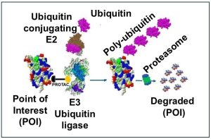
Targeted Protein Degradation (TPD) therapies, including PROTACs (Proteolysis-Targeting Chimeras) and molecular glues, represent a transformative approach in drug development. These therapies leverage the body’s natural protein degradation machinery, such as the ubiquitin-proteasome system, to selectively eliminate disease-causing proteins. While TPD drugs hold immense promise, particularly for targeting “undruggable” proteins in diseases like cancer, their development requires robust Good Manufacturing Practice (GMP) potency assays to ensure safety, efficacy, and regulatory compliance.
Targeted protein degradation (TPD) employs diverse strategies to eliminate specific proteins, offering innovative therapeutic approaches. Key methods include PROTACs, which use bifunctional molecules to link target proteins to E3 ubiquitin ligases for proteasomal degradation, and molecular glues, which induce protein-protein interactions for ubiquitination. LYTACs and GlueTACs target extracellular or membrane proteins for lysosomal degradation, while AUTACs, ATTECs, and MADTACs utilize autophagy pathways. AbTACs employ antibodies for targeted degradation, and dTAGs, HyT, and degron tags are valuable in research applications. Other techniques, such as SNIPERs, IMiDs, and bispecific aptamer chimeras, focus on specialized degradation pathways, demonstrating the versatility and precision of TPD in addressing previously undruggable targets. New technologies, such as dual-action degrader antibody conjugates (DACs), are currently in progress. Details of various TPD technologies are available on our Targeted Protein Degradation Services page.
This article explores the challenges and strategies for developing GMP-compliant potency assays for PROTACs and other TPD drugs, focusing on their unique mechanisms of action (MOA), regulatory requirements, and the transition from research-grade assays to GMP-ready methods.
Importance of MOA and Potency Assays in TPD Drug Development
Understanding the Mechanism of Action (MOA) and developing robust potency assays are critical for the successful development of targeted protein degraders, such as PROTACs and molecular glues. MOA studies guide the design of assays that measure the drug’s interaction with its target, confirm the intended degradation mechanism, and minimize off-target effects. This knowledge defines the biological target and therapeutic goals, forming the foundation for effective drug development.
Potency assays are essential for demonstrating a drug’s efficacy by accurately measuring its ability to achieve the desired therapeutic effect. They provide data for optimizing drug candidates, predicting clinical outcomes, and comparing compounds. The synergy between MOA understanding and potency assay development is vital: MOA insights inform assay design, while potency assays reveal nuances in the MOA. This interplay accelerates the translation of targeted protein degraders into effective therapies by streamlining drug optimization, improving preclinical predictions, and supporting informed decision-making.
For Targeted Protein Degradation (TPD) drugs, potency assays must specifically measure the drug’s ability to degrade the target protein and validate the MOA, which involves forming a ternary complex (target protein, E3 ligase, and degrader) and subsequent ubiquitination and degradation. These assays are critical for regulatory approval, as they demonstrate the drug’s biological activity and therapeutic potential.
Regulatory Definitions of Potency
US FDA (1):
“Potency is defined as “the specific ability or capacity of the product, as indicated by appropriate laboratory tests or by adequately controlled clinical data obtained through the administration of the product in the manner intended, to effect a given result.” (21 CFR 600.3(s)). Strength is defined as “[t]he potency, that is, the therapeutic activity of the drug product as indicated by appropriate laboratory tests or by adequately developed and controlled clinical data. . . .” (21 CFR 210.3(b)(16)). Regulations require that “[t]ests for potency shall consist of either in vitro or in vivo tests, or both, which have been specifically designed for each product so as to indicate its potency in a manner adequate to satisfy the interpretation of potency given by the definition in § 600.3(s) of this chapter.” (21 CFR 610.10).”
ICH Q6B Specifications for Biotechnological Products (2)
“Potency (expressed in units) is the quantitative measure of biological activity based on the attribute of the product which is linked to the relevant biological properties, whereas, quantity (expressed in mass) is a physicochemical measure of protein content. Mimicking the biological activity in the clinical situation is not always necessary. A correlation between the expected clinical response and the activity in the biological assay should be established in pharmacodynamic or clinical studies. “
Why Common MOA and Potency Assays May Not Be Suitable for GMP Assays in Regulatory Approvals
Common Mechanism of Action (MOA) and potency assays, although crucial in early-stage research, often fall short of the stringent requirements needed for Good Manufacturing Practice (GMP) assays in regulatory approvals, such as those mandated by the FDA. One significant limitation lies in their lack of standardization and reproducibility. Research assays are inherently exploratory and flexible, which can lead to variability in results. In contrast, GMP assays demand rigorous standardization to ensure consistency across batches, laboratories, and time points, as stipulated by FDA regulations (21 CFR 610.10). Additionally, early-stage assays often lack formal validation, failing to meet the requirements for accuracy, precision, specificity, linearity, and robustness that are critical for GMP compliance. Regulatory agencies, such as the FDA and ICH, require validated assays to guarantee reliability in assessing product quality (ICH Q2(R2)).
Furthermore, many MOA assays, such as Western blotting and immunoprecipitation, are qualitative or semi-quantitative, and therefore do not meet the quantitative rigor required for GMP assays. GMP assays must deliver precise metrics like EC50 or DC50 values, which correlate with clinical efficacy and safety (FDA, 21 CFR 600.3(s)). The reagents used in MOA assays also pose a challenge, as they are often research-grade and lack the traceability and quality assurance required under GMP standards. GMP assays, on the other hand, necessitate regulatory-compliant reagents with detailed documentation, such as certificates of analysis (ICH Q6B).
Additionally, the complex experimental designs of advanced techniques like AlphaLISA, NanoBRET, or mass spectrometry, while powerful, are impractical for routine GMP testing. GMP assays must be simplified, scalable, and robust enough for use in quality control environments. MOA assays typically focus on general mechanistic validation, whereas GMP assays must directly link to critical quality attributes (CQAs) like consistency, efficacy, and stability. Regulatory submissions require extensive documentation, including protocols, validation reports, and standard operating procedures (SOPs), which MOA assays often lack (FDA Guidance for Industry, 2011).
Lastly, scalability and cost-effectiveness present another hurdle. GMP assays must be high-throughput and economical, while many MOA assays are labor-intensive and reliant on specialized equipment. Regulatory guidelines, such as ICH Q2(R2), emphasize risk mitigation for product quality and patient safety, areas where MOA assays are insufficiently designed to excel. In summary, while MOA and potency assays play a vital role in early development, transitioning to GMP assays is essential to meet regulatory standards. This requires tailored methods that prioritize standardization, validation, and a focus on product quality and safety (FDA, 21 CFR 210.3(b)(16); ICH Q6B).
Challenges in Developing GMP Potency Assays for TPD Drugs
Developing GMP potency assays for PROTACs and other targeted protein degradation (TPD) drugs poses several unique challenges. One major hurdle is the complex mechanisms of action (MOA) of TPD drugs, which involve multi-step processes such as target engagement, ternary complex formation, ubiquitination, and proteasomal degradation. Capturing these steps within a single assay is difficult, and differentiating between on-target degradation and off-target effects further complicates potency evaluation. Additionally, many target proteins exist in low abundance, requiring highly sensitive detection methods like mass spectrometry or quantitative immunoassays. Cellular variability presents another challenge, as differences in E3 ligase expression, proteasome activity, and target protein levels across cell lines can affect TPD drug efficacy, necessitating careful model selection and rigorous controls. Unlike traditional small-molecule drugs, PROTACs act catalytically, with a single molecule capable of degrading multiple target proteins. This catalytic nature complicates traditional pharmacokinetic/pharmacodynamic (PK/PD) assessments. Early-stage research assays, such as Western blotting or immunoprecipitation, often lack the standardization and reproducibility required for GMP compliance. Furthermore, regulatory requirements mandate that GMP assays be rigorously validated for accuracy, precision, specificity, linearity, and robustness. These assays must also directly link to the drug’s critical quality attributes (CQAs), such as consistency, efficacy, and stability, adding another layer of complexity to assay development.
Key Considerations for GMP Potency Assay Development
To overcome the challenges in developing GMP potency assays for TPD drugs, several key considerations must be addressed. Standardization and reproducibility are critical, requiring GMP assays to deliver consistent results across different batches, laboratories, and time points. This involves clearly defining critical reagents, experimental conditions, and acceptance criteria. Quantitative and sensitive detection methods, such as ELISA, mass spectrometry, flow cytometry, cellular immunoassays, or cell based reporter assays should be employed to measure target protein degradation and its downstream functional effects accurately. Validation of assay parameters is another essential step, as GMP assays must demonstrate accuracy, precision, specificity, linearity, and robustness to meet regulatory requirements and ensure reliability in batch release testing. Additionally, potency assays must not only quantify target protein degradation but also establish a clear linkage to biological outcomes, such as changes in gene expression, cell viability, or signaling pathways. Scalability and high-throughput capability are equally important; GMP assays must be streamlined and optimized for efficient testing in quality control environments, incorporating simplified experimental designs and regulatory-compliant reagents to ensure practicality and compliance.
Transitioning from Research-Grade to GMP-Compliant Assays
The transition from research-grade assays to GMP-compliant potency assays involves several stages (see also Fig. 1):
- Development Stage: During preclinical research, the initial assay is designed to represent the product’s mechanism of action (MOA) under a limited set of conditions.
- Optimization Stage: As the product enters early clinical trials, the assay is prepared for expanded use in a commercial setting. This involves defining specifications for critical reagents and considering the availability of materials and specialized equipment.
- Qualification/Validation Stage: Prior to Phase 3 trials or before submitting Biologics License Application (BLA) or Marketing Authorization Application (MAA), the assay undergoes protocol-driven evaluation of reproducibility, accuracy, precision, specificity, robustness, and range. This stage is crucial for setting acceptance criteria for the subsequent validation.
Fig. 1: Stages of TPD potency assay development process.

Examples of GMP Potency Assays for TPD Drugs
Strategies for developing potency assays for targeted protein degradation (TPD) drugs, such as PROTACs, involve assessing key aspects of their mechanism of action. Commonly used methods include cytotoxicity assays, flow cytometry, ELISA (enzyme-linked immunosorbent assay), cell-based assays, and reporter assays. Some examples are shown in Table 1. Details of potency assays for PROTACs are described in the article: Advancing PROTAC Therapeutics: Potency Assays for FDA Compliance.
Table 1: Examples of MOA and Potency assays for PROTAC and Molecular Glue Targets
| Target Protein | MOA Assays | Potency Assays | Examples |
|---|---|---|---|
| BRD4 (Bromodomain) (3, 4) | Western blot, NanoBRET | MYC reporter assays, gene expression (MYC, BCL2) | BET degraders like dBET1 |
| IRAK4 (4) | Ubiquitination assay, ALPHA assay | NF-κB activity, cytokine release (IL-6, TNF-α) | PROTACs for NF-κB signaling |
| BCL-XL (5) | Annexin V staining, flow cytometry for apoptosis | Caspase activity, cleaved PARP ELISA | Dual degraders for BCL-XL and BCL-2 |
| EZH2 (Methyltransferase) (6) | Histone methylation via ELISA | Gene expression (CDKN1A), qPCR | EZH2-targeted PROTACs |
Phase-Appropriate Potency Assays for TPD Drugs
The development of a potency assay follows a phase-appropriate strategy aligned with the therapeutic product’s lifecycle. During preclinical research, initial assays are designed to reflect the drug’s mechanism of action (MOA) under controlled conditions. As the product advances to early clinical trials, the assay is optimized for broader use, focusing on scalability, reproducibility, and defining critical reagents. Prior to Phase 3 trials or regulatory submissions, the assay undergoes rigorous qualification and validation to ensure accuracy, precision, specificity, and robustness, meeting regulatory standards for Good Manufacturing Practice (GMP). This phased approach ensures that potency assays evolve alongside the drug’s development, providing reliable data for lot release, stability testing, and regulatory approvals while maintaining alignment with the product’s critical quality attributes (CQAs). An example of the phase-specific potency assays for PROTACs/molecular glues is shown in the Table in Appendix 3.
Regulatory Considerations for cGMP Potency Assays
Regulatory agencies, such as the FDA and EMA, require current GMP (cGMP) potency assays to meet stringent standards, including:
• Direct Link to CQAs: Assays must demonstrate consistency, efficacy, and stability of the drug product.
• Comprehensive Documentation: Detailed protocols, validation reports, and standard operating procedures (SOPs) are required for regulatory compliance.
• Risk Mitigation: Assays must address potential risks to product quality, efficacy, and patient safety, as outlined in guidelines like ICH Q2(R2) and FDA bioassay guidance.
Conclusion
Developing GMP potency assays for PROTACs and other TPD drugs is a complex but essential process for ensuring the safety, efficacy, and regulatory compliance of these innovative therapies. By addressing challenges such as assay standardization, sensitivity, and linkage to biological outcomes, researchers can create robust GMP assays that meet regulatory requirements and unlock the full potential of targeted protein degradation in treating diseases like cancer. As the field of TPD continues to evolve, advancements in assay development will play a pivotal role in bringing these groundbreaking therapies to patients.
References
- 1. Guidance for Industry Potency Tests for Cellular and Gene Therapy Products
- 2. ICH Topic Q 6 B Specifications: Test Procedures and Acceptance Criteria for Biotechnological/Biological Products
- 3. Bromodomain and extraterminal (BET) proteins: biological functions, diseases and targeted therapy
- 4. Targeted protein degradation: advances in drug discovery and clinical practice
- 5. Control of apoptosis by the BCL-2 protein family: implications for physiology and therapy
- 6. Degraation of Polycomb Repressive Complex 2 with an EED-Targeted Bivalent Chemical Degrader
- 7. A Tale of Two Tails: Efficient Profiling of Protein Degraders by Specific Functional and Target Engagement Readouts
- 8. Affinity and cooperativity modulate ternary complex formation to drive targeted protein degradation
- 9. SPR-Measured Dissociation Kinetics of PROTAC Ternary Complexes Influence Target Degradation Rate
- 10. Assays and technologies for developing proteolysis targeting chimera degraders
- 11. PO-449 Optimisation of an AlphaLISA assay for the characterisation of PROTAC-induced ternary complexes within cell lysates
- 12. General Stepwise Approach to Optimize a TR-FRET Assay for Characterizing the BRD/PROTAC/CRBN Ternary Complex
- 13. A proximity biotinylation-based approach to identify protein-E3 ligase interactions induced by PROTACs and molecular glues
- 14. Design of a Cereblon construct for crystallographic and biophysical studies of protein degraders
- 15. The In-Cell Western immunofluorescence assay to monitor PROTAC mediated protein degradation
- 16. Degradome analysis to identify direct protein substrates of small-molecule degraders
- 17. Tracking the PROTAC degradation pathway in living cells highlights the importance of ternary complex measurement for PROTAC optimization
- 18. A Shift in Thinking: Cellular Thermal Shift Assay-Enabled Drug Discovery
- 19. The Advantages of Targeted Protein Degradation Over Inhibition: An RTK Case Study
- 20. Accelerating PROTAC drug discovery: Establishing a relationship between ubiquitination and target protein degradation
- 21. Quantitative Live-Cell Kinetic Degradation and Mechanistic Profiling of PROTAC Mode of Action
- 22. Monitoring and deciphering protein degradation pathways inside cells
- 23. Functional Genomics Identify Distinct and Overlapping Genes Mediating Resistance to Different Classes of Heterobifunctional Degraders of Oncoproteins
- 24. Flexible Fitting of PROTAC Concentration-Response Curves with Changepoint Gaussian Processes
- 25. Three-Dimensional Cell Cultures in Drug Discovery and Development
- 26. A cyclic peptide-based PROTAC induces intracellular degradation of palmitoyltransferase and potently decreases PD-L1 expression in human cervical cancer cells
- 27. New-generation advanced PROTACs as potential therapeutic agents in cancer therapy
Related Articles/Blogs
Development of Cell-Based Potency Assays: Case Studies and Blogs from Marin Biologic Laboratories (MarinBio)




Our Recent Publication/Presentation
American Society of Hematology (ASH) Annual Meeting 2024.
Appendix
Appendix Table 1: Common MOA Assays for PROTACs and Molecular Glues
| Category | Assay Name | Purpose | Key Techniques/Notes |
|---|---|---|---|
| Target Engagement Assays | Thermal Shift Assays (TSA) (7) | Detects stabilization of the target protein upon binding. | Measures melting temperature shifts. |
| Isothermal Titration Calorimetry (8) | Quantifies binding affinity. | Provides thermodynamic data. | |
| Surface Plasmon Resonance (SPR) (9) | Measures real-time binding kinetics. | Useful for studying dynamic interactions. | |
| Fluorescence Polarization (FP) (10) | Detects ligand binding to the protein target. | Sensitive to small molecule interactions. | |
| Ternary Complex Formation | AlphaLISA (11) | Confirms proximity between the target protein and E3 ligase. | Homogeneous, high-throughput technique. |
| FRET/BRET (12) | Detects ternary complex formation in real time. | Useful for live-cell studies. | |
| Proximity Ligation Assay (PLA) (13) | Visualizes ternary complex formation in cells. | Requires specific antibodies for detection. | |
| Cryo-EM/X-ray Crystallography (14) | Provides structural insights into the ternary complex. | High-resolution but resource-intensive. | |
| Protein Degradation Assays | Western Blotting (15) | Detects changes in target protein levels. | Semi-quantitative. |
| ELISA (15) | Uses antibodies to detect the target protein. | High sensitivity and specificity. | |
| Quantitative Mass Spectrometry (16) | Measures proteome-wide changes in protein levels. | Highly quantitative and sensitive. | |
| Live Cell Assays (17) | Monitors real-time degradation in live cells. | Combines degradation kinetics with live-cell monitoring. | |
| Cellular Thermal Shift Assay (CETSA) (18) | Detects changes in thermal stability of the degraded protein. | Confirms degradation in live cells. | |
| Ubiquitination Assays | Immunoprecipitation (IP) (15) | Pulls down the target protein and detects ubiquitin conjugates. | Confirms ubiquitination. |
| Mass Spectrometry (16) | Identifies ubiquitinated residues. | High resolution, allows site mapping. | |
| Proteasome Assays (19) | Confirms ubiquitination in the presence of proteasome inhibitors (e.g., MG132). | Differentiates ubiquitination from degradation. | |
| Functional Assays | Reporter Gene Assays (20) | Measures downstream effects on gene expression. | Detects functional consequences of degradation. |
| Cell Viability Assays (21) | Assesses therapeutic efficacy (e.g., MTT, CellTiter-Glo). | Common in cancer models. | |
| Pathway Analysis (22) | Detects changes in signaling pathways affected by protein degradation. | Includes phosphoproteomics and ELISA. | |
| Selectivity Assays | Proteomics (TMT or SILAC) (16) | Detects off-target effects across the proteome. | Highly quantitative. |
| CRISPR Knockout/RNAi (23) | Confirms on-target degradation effects. | Validates target specificity. | |
| Degron Mutant Studies (24) | Uses mutations in degron sequences to confirm specificity. | Specific for target proteins. | |
| E3 Ligase-Dependent Assays | E3 Ligase Knockdown/Knockout (23) | Assesses dependency on specific E3 ligases. | Uses siRNA or CRISPR. |
| Mutant E3 Ligase Studies (23) | Confirms degradation by testing inactive or mutant ligases. | Validates E3 ligase involvement. |
Appendix Table 2: Common Potency Assays for PROTACs and Molecular Glues
| Category | Assay Name | Purpose | Key Techniques/Notes |
|---|---|---|---|
| Target Protein Degradation | Western Blotting (15) | Quantifies the level of target protein degradation. | Semi-quantitative and widely used for initial potency evaluation. |
| NanoBRET (10) | Monitors real-time degradation of tagged target proteins in live cells. | Provides degradation kinetics in a high-throughput format. | |
| Cellular Thermal Shift Assay (CETSA) (7) | Confirms degradation by detecting thermal stability changes of the target protein. | Useful for cellular contexts. | |
| High-Throughput ELISA (10) | Measures the reduction of target protein levels in cell lysates. | Ideal for high-throughput screening of multiple compounds. | |
| Functional Activity | Reporter Gene Assays (20) | Detects downstream functional effects of target degradation on gene expression. | Indicates functional efficacy in biological pathways. |
| Cell Viability Assays (21) | Evaluates the impact of degradation on cell proliferation or survival (e.g., MTT, CellTiter-Glo). | Common for assessing anti-cancer activity. | |
| Signaling Pathway Assays (22) | Monitors changes in phosphorylation or other signaling events downstream of protein degradation. | Uses phospho-specific antibodies or proteomics approaches. | |
| Selectivity and Specificity | Proteomics (TMT or SILAC) (16) | Quantifies proteome-wide effects to confirm selectivity of the compound. | Useful for identifying off-target degradation. |
| CRISPR Knockout/RNAi (23) | Confirms dependency of observed effects on the target protein. | Validates on-target potency. | |
| Mutant Target Studies (24) | Tests degradation-resistant mutants to confirm specificity. | Ensures degradation is specific to the target. | |
| Dose-Response Analysis | Dose-Response Degradation Curves (DC50) (25) | Determines the concentration of the compound required for 50% degradation of the target protein. | Provides key potency metrics (e.g., DC50). |
| Hill Slope Analysis (10) | Evaluates the steepness of the dose-response curve for degradation. | Helps in understanding cooperativity or potency of the compound. | |
| Time-Dependent Degradation (Dmax) (10) | Measures the maximum degradation achievable and the time required to reach it. | Indicates the efficiency and kinetics of the degrader. | |
| Ubiquitination-Related | Ubiquitination Assays (15-16) | Confirms target protein ubiquitination before degradation. | Immunoprecipitation or proteasome inhibitor studies can complement potency assessments. |
| Proteasome Inhibition Potency (17) | Evaluates the buildup of ubiquitinated protein in the presence of proteasome inhibitors. | Indicates if degradation is proteasome-dependent. | |
| Cellular Models | 2D and 3D Cellular Assays (26) | Measures potency in simple and complex cellular environments. | 3D models better mimic in vivo conditions. |
| Co-Culture Assays (27) | Evaluates potency in mixed cell populations to assess specificity and functional outcomes. | Useful for cancer models or immune-oncology studies. | |
| Animal Models | Biomarker Analysis in Vivo (28) | Measures the reduction of target protein or functional biomarkers in tissues or plasma. | Useful for assessing in vivo potency. |
| Pharmacokinetic/Pharmacodynamic (PK/PD) (10) | Correlates compound exposure with target degradation and functional effects in vivo. | Helps translate potency from in vitro to in vivo systems. |
Appendix Table 3: Phase-Appropriate Potency Assays for TPD Drugs
| Clinical Phase | Objective | Assays/Methods |
|---|---|---|
| Preclinical and Early Phase (Phase I) | Demonstrate target engagement, degradation efficiency, and preliminary safety | In vitro target degradation assays (e.g., Western blotting, ELISA, flow cytometry) |
| Demonstrate target engagement, degradation efficiency, and preliminary safety | Biochemical assays for purified protein systems | |
| Demonstrate target engagement, degradation efficiency, and preliminary safety | In vivo PK/PD studies and xenograft models | |
| Mid-Stage (Phase II) | Establish efficacy, dose optimization, and safety in larger patient populations | Broad target degradation assays in primary or patient-derived cells |
| Establish efficacy, dose optimization, and safety in larger patient populations | Functional readout assays to link degradation with biological effects | |
| Establish efficacy, dose optimization, and safety in larger patient populations | Biomarker assays (pharmacodynamic or disease-specific) | |
| Establish efficacy, dose optimization, and safety in larger patient populations | Resistance mechanism assays to identify resistance pathways | |
| Establish efficacy, dose optimization, and safety in larger patient populations | Drug stability, bioavailability, immunogenicity, and toxicity assays | |
| Late-Stage (Phase III) | Confirm efficacy and safety, and validate assays for regulatory approval and manufacturing | Validated potency assays for batch-to-batch consistency (e.g., mass spectrometry) |
| Confirm efficacy and safety, and validate assays for regulatory approval and manufacturing | Pharmacokinetic/pharmacodynamic (PK/PD) correlation for dose-response relationships | |
| Confirm efficacy and safety, and validate assays for regulatory approval and manufacturing | Mechanistic assays under physiologically relevant conditions | |
| Confirm efficacy and safety, and validate assays for regulatory approval and manufacturing | Clinical endpoint assessments (ORR, PFS, OS) | |
| Commercialization (Post-Approval) | Ensure long-term consistency, stability, and regulatory compliance | GMP-compliant lot release assays |
| Ensure long-term consistency, stability, and regulatory compliance | Stability monitoring and post-marketing surveillance assays |
With over 30 years of expertise in cell-based potency assay development, we deliver precise, GMP-compliant potency assays for PROTACs, molecular glues, and other TPD drug candidates to accelerate your clinical success.
Click here to learn more about Targeted Protein Degradation Services
Targeted Protein Degradation (TDP) Assay Services Offered by MarinBio
Target Engagement Assays
These assays measure the binding affinity of the degrader to the target protein and the E3 ligase.
- Fluorescence Polarization (FP) Assays: Used to determine the binding affinity of the degrader to the target protein or E3 ligase by measuring changes in fluorescence polarization.
- Flow Cytometry: Used to measure binding affinity of degraders to target proteins or E3 ligases in live cells.
Mechanism of Action (MOA) Assays
MOA assays provide a comprehensive understanding of the TPD mechanism, from initial target engagement to final protein degradation, helping researchers optimize and characterize TPD compounds throughout the drug discovery process.
- Ternary Complex Formation Assays: Co-immunoprecipitation (Co-IP).
- Ubiquitination Assays: In vitro ubiquitination assays and Western blotting for ubiquitinated proteins.
- Cellular Degradation Assays: ELISA, Western blot, qPCR
- Functional Cellular Assays: Cell viability assays, Reporter gene assays
- Cell-Free Degradation Assays: In vitro ubiquitination Assays
- Flow Cytometry: Real-time monitoring of protein degradation kinetics in live cells, measuring ubiquitination of target proteins.
Selectivity Assays
- Off-Target Screening: Identify and quantify potential off-target effects of the degrader.
- Cytokine Release Assays: Measures the release of cytokines to assess the immunomodulatory effects of the degrader.
Potency Assays
A potency assay measures the effectiveness of a degrader molecule in degrading a target protein. These assays determine the minimum compound amount needed for a biological response, crucial for drug discovery and development. The following assays can be used:
ADME/Tox Studies
ADME/Tox assays provide crucial data for predicting the pharmacokinetic and pharmacodynamic properties and evaluating the safety profile of TPD compounds. See our ADME/Tox service page for more details.
GMP/GLP Services
- Potency Assays
- Stability Testing
- Lot Release Testing
- Serum/plasma-based pharmacokinetic (PK), pharmacodynamic (PD), and toxicology studies.
Custom Stable Cell Line Development
Disclaimer: This information is intended solely for research purposes and does not serve as medical advice.
