


Introduction
T-cell dysfunction is a major barrier to effective and durable immunity in cancer, chronic viral infection, and adoptive cellular therapy. Persistent antigen exposure and prolonged T-cell receptor (TCR) signaling can progressively impair proliferative capacity, cytokine production, cytotoxic activity, metabolic fitness, and memory formation. This process, commonly referred to as T-cell exhaustion, is accompanied by sustained expression of inhibitory receptors such as PD-1, transcriptional and epigenetic remodeling, and profound alterations in cellular metabolism. Although exhaustion initially provides an adaptive mechanism that limits immunopathology during persistent antigen stimulation, progressive differentiation toward terminal exhaustion can severely restrict the ability of T cells to control tumors or chronic infections.
A major conceptual advance has therefore been the recognition that T-cell exhaustion represents a developmental continuum rather than a uniform terminal phenotype. At one end of this continuum are TCF1+ progenitor-exhausted or stem-like T cells (T-PEX) that retain proliferative and self-renewal capacity and can continuously generate more differentiated effector populations. At the opposite end are terminally exhausted T cells characterized by limited proliferative potential, altered metabolism, extensive epigenetic remodeling, and high expression of inhibitory receptors. The therapeutic objective has changed from simply attempting to reactivate dysfunctional cells toward preserving the populations capable of sustaining long-term immunity.
TCF1⁺ progenitor-exhausted T cells are a subset of chronically stimulated CD8⁺ T cells that retain stem-like properties, including self-renewal and proliferative capacity, despite expressing exhaustion-associated markers such as PD-1. They serve as a renewable reservoir that can generate more differentiated effector and terminally exhausted T cells. T-PEX cells are particularly important therapeutically because they provide a major source of T-cell expansion following PD-1 checkpoint blockade. In cancer, many TCF1⁺ progenitor-exhausted (T-PEX) cells are tumor-antigen specific, particularly the populations that sustain antitumor responses and respond to checkpoint blockade. Preserving and expanding this population is therefore emerging as an important strategy for achieving durable antitumor immunity.
Recent studies have begun to define the molecular architecture underlying this differentiation process. TCF1, MYB, PD-1, TOX, and NR4A-family transcription factors constitute an important framework linking progenitor maintenance with progression toward terminal dysfunction. TCF1 identifies and contributes to maintenance of the stem-like state, while MYB has emerged as an important regulator of stem-like tumor-reactive T cells within tumor-draining lymph nodes. These lymphoid populations can function as reservoirs that continually replenish tumor-infiltrating T cells. PD-1 occupies a more complex position than originally appreciated: although PD-1 inhibits TCR signaling and is an established therapeutic target, recent evidence indicates that controlled PD-1 signaling can also restrain excessive activation and contribute to maintenance of high-avidity stem-like T cells during persistent antigen exposure.
Conversely, sustained TCR signaling and persistent NFAT activity promote transcriptional programs involving TOX and NR4A-family proteins. TOX and NR4A are transcription factors that help establish and maintain the dysfunctional state of chronically stimulated T cells. They are particularly important during persistent antigen exposure in cancer and chronic infection, where sustained TCR signaling activates NFAT-dependent transcriptional programs. These discoveries have helped explain why simply removing an inhibitory receptor signal may be insufficient to completely restore T-cell function. Once exhaustion-associated transcriptional and epigenetic programs become established, the dysfunctional phenotype can become increasingly resistant to reversal.
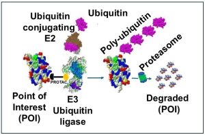
Recent studies revealed that T-cell fate is regulated not only through transcription and checkpoint signaling but also through proteostasis, mitochondrial dynamics, oxidative-stress responses, nutrient sensing, and ion-channel activity. Particularly important examples include pathways involving KLHL6, PTGIR axis, and NFAT1. KLHL6 is an adaptor for an E3 ubiquitin ligase complex that protects CD8⁺ T cells from dysfunction by regulating protein degradation and mitochondrial fitness. KLHL6 promotes ubiquitination and degradation of TOX, thereby limiting accumulation of exhaustion-associated transcriptional programs. During chronic stimulation, loss or reduction of KLHL6 contributes to TOX accumulation, mitochondrial dysfunction, and progression toward terminal exhaustion. Increasing KLHL6 activity or expression may therefore provide a strategy for generating more persistent, exhaustion-resistant antitumor T cells. The identification of PTGIR as a regulator of terminal exhaustion identifies a potentially druggable GPCR within the exhaustion network. Similarly, the discovery that methionine availability regulates Ca²⁺ signaling and NFAT1 activity provides a mechanistic connection between nutrient availability during T-cell activation and subsequent commitment to dysfunctional states.
Together, these discoveries support a substantially broader model of T-cell dysfunction in which cellular differentiation, inhibitory signaling, transcriptional regulation, epigenetic state, proteostasis, metabolism, and the extracellular microenvironment are tightly interconnected. This expanded understanding is also creating new therapeutic opportunities. Checkpoint inhibitors can be combined with approaches that preserve TCF1+ progenitor populations, manipulate cytokine or costimulatory signaling, alter exhaustion-associated transcriptional programs, restore mitochondrial fitness, or engineer CAR-T and TCR-T cells that are intrinsically resistant to dysfunctional differentiation.
Molecular Framework and Emerging Therapeutic Targets in T-Cell Dysfunction
A major advance in understanding T-cell dysfunction has been the recognition that exhausted CD8+ T cells are not a homogeneous population but instead occupy a differentiation continuum extending from T-PEX cells to progressively differentiated and terminally exhausted T cell populations. This framework has important therapeutic implications because the capacity for proliferation, effector-cell generation and response to immune checkpoint blockade (ICB) is concentrated disproportionately within the progenitor compartment rather than terminally exhausted cells. Recent studies have further refined this model by identifying spatial, transcriptional, proteostatic and metabolic mechanisms controlling progression through these states.
TCF1: maintaining the stem-like progenitor compartment
T-cell factor 1 (TCF1), is a defining regulator of stem-like/progenitor exhausted CD8+ T cells. TCF1+PD-1+ tumor-reactive T cells retain proliferative potential, self-renewal properties and the ability to generate more differentiated TCF1- effector/exhausted progeny. Importantly, these cells provide a major proliferative response to checkpoint blockade, whereas terminally differentiated TCF1- exhausted cells have substantially less capacity for durable expansion.
The therapeutic significance of TCF1 therefore extends beyond its usefulness as a phenotypic marker. The TCF1+ compartment functions as a renewable source of tumor-reactive effector cells. Consequently, therapeutic strategies that preserve TCF1-associated stemness may be more effective than approaches directed exclusively toward reactivating terminally exhausted cells. The importance of anatomical location has also become increasingly apparent. Stem-like T-PEX cells are maintained not only within tumors but also within tumor-draining lymph nodes (tdLNs), where they can continually replenish tumor-infiltrating populations. This observation shifts part of the therapeutic focus away from the tumor itself toward lymphoid niches that maintain tumor-reactive T-cell precursors.
MYB: maintaining lymph-node-derived stem-like T cells
The transcription factor MYB (c-Myb) has recently emerged as an important regulator of this extratumoral progenitor compartment. Recently, researchers demonstrated that MYB-dependent stem-like T-PEX cells residing in tdLNs sustain CD8+ T-cell infiltration into tumors and contribute to the response to ICB. In contrast, intratumoral TCF1+ populations progressively acquire a tissue-residency program that restricts their contribution to systemic replenishment. This distinction suggests that the location and developmental context of a TCF1+ cell may be as important as TCF1 expression itself. MYB appears to participate in maintaining the lymph-node stem-like population capable of repeatedly supplying tumors with differentiated progeny. The same study identified TGF-β as an opposing regulator that promotes tissue residency and restricts the abundance of stem-like T-PEX cells in tdLNs.
The resulting model places TCF1 and MYB at the progenitor-preserving end of the exhaustion spectrum, making maintenance of these programs an attractive therapeutic objective. Manipulation of MYB itself may be difficult because of its broad biological functions, but pathways controlling MYB-dependent T-PEX maintenance could provide opportunities for preserving therapeutically responsive T cells.
TOX : establishing and reinforcing the exhausted state
At the more differentiated end of the spectrum, TOX and NR4A-family transcription factors are major regulators of exhaustion-associated cellular programs.
Persistent TCR signaling activates calcineurin/NFAT-dependent transcriptional pathways that induce TOX. TOX subsequently participates in extensive transcriptional and chromatin remodeling that establishes the characteristic exhausted T-cell state. TOX is therefore not simply another marker of dysfunctional cells; it contributes directly to the development of exhaustion-associated transcriptional and epigenetic identity. Importantly, TOX should not necessarily be considered an exclusively pathological protein. Exhaustion represents an adaptation that permits T cells to survive persistent antigenic stimulation. Complete elimination of TOX-dependent programs can compromise the persistence of chronically stimulated cells. Therapeutically, the objective may therefore be to modulate TOX abundance or activity sufficiently to limit terminal differentiation while preserving the adaptive survival functions of the exhaustion program.
Emerging Therapeutic Nodes
The major advances in treating T-cell dysfunction reflect a shift from attempting to broadly stimulate exhausted T cells toward preserving functional T-cell states, expanding TCF1⁺ stem/progenitor populations, and preventing progression to terminal exhaustion. Therapeutic approaches now target multiple components of the dysfunctional state, including inhibitory receptors, transcriptional and epigenetic programming, metabolic insufficiency, cytokine signaling, costimulation, and the suppressive tumor microenvironment.
Table 1. Major Therapeutic Strategies for Overcoming T-Cell Dysfunction
| Therapeutic strategy | Major targets | Mechanistic rationale | Development status |
|---|---|---|---|
| Preservation/ expansion of TCF1⁺ progenitor-exhausted cells | TCF1/TCF7; PD-1⁺TCF1⁺ CD8 T cells | Maintains a self-renewing population capable of producing differentiated antitumor effectors | Major emerging therapeutic concept; potentially important for durable responses |
| Transcriptional reprogramming | TOX, NR4A, NFAT/AP-1, c-Jun | Chronic antigen signaling establishes transcriptional programs that reinforce exhaustion | Strong preclinical evidence; particularly attractive for engineered T-cell therapies |
| Epigenetic reprogramming | DNMT3A, DNA methylation, chromatin remodeling | Exhaustion-associated chromatin and methylation programs restrict reversibility | Emerging strategy; targeted or cell-intrinsic manipulation may offer advantages over systemic epigenetic therapy |
| Metabolic reprogramming | PGC-1α, mitochondrial biogenesis, ROS, nutrient utilization, mitophagy | Dysfunctional T cells develop mitochondrial impairment and reduced metabolic flexibility in the TME | Particularly promising for solid tumors with metabolically hostile microenvironments |
| Cytokine engineering | IL-2, IL-7, IL-15, IL-21; targeted cytokine variants | Provides survival, proliferative and differentiation signals while attempting to avoid toxicity of systemic cytokines | Rapidly developing; particularly suitable for combination with checkpoint blockade and cellular therapy |
| Restoration of costimulation | CD28, 4-1BB/CD137, OX40, CD27 | Restores positive signaling required for proliferation, survival and differentiation | Established principle in next-generation tumor-targeted agonists under development |
Therapeutic approaches to T-cell dysfunction can be divided into two broad categories. The first attempts to restore the activity of T cells that have already entered dysfunctional states. Immune checkpoint blockade is the best-established example. Blocking PD-1/PD-L1, CTLA-4 or LAG-3 removes inhibitory signals, but the response is strongly influenced by the differentiation state of the responding T cells. Epigenetic studies have shown that terminally exhausted cells retain exhaustion-associated chromatin programs even after PD-1 blockade, helping explain why checkpoint inhibition does not completely restore conventional memory T-cell function.
The second, and increasingly important strategy, attempts to prevent or delay terminal dysfunction. Identification of TCF1⁺ progenitor-exhausted CD8 T cells has been particularly important to this concept. These cells maintain proliferative and self-renewal potential and can generate more differentiated tumor-reactive progeny. Rather than forcing terminally exhausted cells back into a functional state, therapies may therefore achieve greater durability by maintaining a renewable TCF1⁺ population and controlling its differentiation.
The emerging model suggests that T-cell dysfunction should not be treated as a single reversible state. Instead, exhaustion represents a differentiation trajectory extending from TCF1⁺ stem/progenitor populations through transitional states to terminally dysfunctional T cells. The therapeutic responsiveness of these populations differs substantially.
Accordingly, the field is moving from a predominantly “reverse exhaustion” model toward a “preserve stemness and prevent terminal exhaustion” model. Checkpoint blockade can release inhibitory signaling, but durable responses may require maintenance of TCF1⁺ progenitor populations capable of continuously supplying functional effector cells. Cytokine and costimulatory signals can then regulate their expansion and differentiation, while metabolic and epigenetic interventions may prevent premature loss of cellular fitness.
This model also provides a rationale for rational combination therapy. Rather than combining immunotherapies solely to increase the magnitude of T-cell activation, future approaches can target different stages of the dysfunctional differentiation pathway. For example, checkpoint blockade to relieve inhibition, IL-2-family cytokine engineering to expand responsive populations, metabolic interventions to maintain cellular fitness, and transcriptional or epigenetic engineering to reduce progression toward terminal exhaustion. Together, these advances represent a transition from nonspecific enhancement of T-cell activation toward deliberate control of T-cell differentiation and long-term functional state.
Conclusion and Future Perspectives
Recent advances have fundamentally changed the understanding of T-cell dysfunction. Exhaustion is no longer adequately described as a uniform state characterized principally by inhibitory-receptor expression and reduced effector function. Instead, it represents a dynamic differentiation process involving distinct cellular populations with markedly different capacities for self-renewal, proliferation, effector generation, and therapeutic responsiveness. Recognition of this heterogeneity provides an important explanation for both the remarkable success and the limitations of current immunotherapies.
Central to this revised model is the distinction between TCF1+ stem/progenitor exhausted T cells and terminally exhausted T cells. The former retain the capacity to proliferate and generate differentiated progeny and constitute an important cellular reservoir supporting responses to checkpoint blockade. The identification of MYB-dependent stem-like populations within tumor-draining lymph nodes further demonstrates that durable antitumor immunity depends not only on cells residing within the tumor but also on lymphoid niches capable of continuously supplying tumor-reactive T cells. Preservation of these populations may therefore be as important therapeutically as reactivation of existing tumor-infiltrating lymphocytes.
At the same time, the role of TOX, and other regulators illustrate the complexity of therapeutically manipulating exhaustion. These molecules should not be viewed exclusively as pathological regulators that must be eliminated. PD-1 can restrain excessive TCR signaling and may contribute to preservation of stem-like cells under conditions of persistent antigen exposure, while TOX-dependent programs can permit chronically stimulated T cells to survive despite simultaneously establishing features of exhaustion. Consequently, permanent genetic deletion of inhibitory or exhaustion-associated pathways may have biological consequences that differ substantially from transient pharmacological modulation. The challenge is therefore to control the intensity, timing, and cellular context of these pathways rather than simply eliminate them.
The recent identification of pathways involving KLHL6, PTGIR, and NFAT1 considerably broadens the therapeutic landscape. KLHL6 establishes proteostasis as an important determinant of T-cell fate by connecting ubiquitin-dependent regulation of TOX with mitochondrial homeostasis. The PTGIR pathway demonstrates that oxidative-stress responses can actively promote terminal exhaustion and, importantly, identifies a cell-surface GPCR that may provide a pharmacologically accessible intervention point. The NFAT1 pathway further demonstrates that nutrient availability during early T-cell activation can alter Ca²⁺ signaling and establish long-lasting consequences for subsequent differentiation. Together, these discoveries connect environmental conditions encountered by T cells directly to the transcriptional machinery governing exhaustion.
These findings also highlight a critical principle for therapeutic development: maximal T-cell activation is not necessarily equivalent to optimal T-cell function. Excessive or sustained signaling can accelerate differentiation, metabolic stress, and loss of proliferative potential. Effective therapies will therefore need to balance immediate cytotoxic activity with preservation of long-term cellular fitness. An important future challenge will be determining which interventions should be combined and at what stage of T-cell differentiation they should be applied. The pathways that preserve a progenitor cell may differ from those required to generate an effective cytotoxic effector, and interventions beneficial during ex vivo expansion may not be appropriate after tumor infiltration. Biomarkers capable of distinguishing T-PEX, transitional, effector-like, and terminal exhausted T cell populations will therefore be essential for matching therapies to specific dysfunctional states.
Ultimately, the emerging objective is not simply to “reverse T-cell exhaustion.” A more realistic and potentially more effective therapeutic strategy is to manage the trajectory of T-cell differentiation, maintaining a renewable pool of stem/progenitor cells while permitting controlled production of metabolically competent cytotoxic effectors and preventing premature commitment to terminal dysfunction. The convergence of checkpoint biology with recent discoveries in transcriptional regulation, proteostasis, metabolism, redox signaling, and nutrient sensing provides an increasingly integrated framework for achieving this goal.
Thus, the field is transitioning from reactivation of exhausted T cells toward deliberate control of T-cell state and fate. If recent mechanistic advances can be translated into selective pharmacologic interventions and rationally engineered cellular therapies, the ability to preserve durable T-cell immunity while maintaining potent antitumor activity may represent one of the most important next steps in cancer immunotherapy and the treatment of other diseases characterized by chronic T-cell dysfunction.
References
- Wu Y, et al. Revitalizing T cells: breakthroughs and challenges in overcoming T cell exhaustion. Signal Transduct Target Ther. 2026. PMID: 41484095.
- T-cell exhaustion in tumor immunology: mechanisms, heterogeneity, and therapeutic strategies. Front Immunol. 2026. PMID: 42220497. doi:10.3389/fimmu.2026.1841281.
- T cell exhaustion landscapes and therapeutic modulation in cancer immunity. 2026. PMID: 42222335.
- T Cell Exhaustion in Cancer Immunotherapy: Heterogeneity, Mechanisms, and Therapeutic Opportunities. Adv Sci (Weinh). 2026. PMID: 41527444.
- Targeting the fate of exhausted CD8+ T cells. Trends Immunol. 2026. PMID: 42538164. doi:10.1016/j.it.2026.07.002.
- Siddiqui I, Schaeuble K, Chennupati V, et al. Intratumoral Tcf1+PD-1+CD8+ T cells with stem-like properties promote tumor control in response to vaccination and checkpoint blockade immunotherapy. Immunity. 2019;50:195-211.e10. PMID: 30635237.
- Prokhnevska N, et al. Lymph-node-derived stem-like but not tumor-tissue-resident CD8+ T cells fuel anticancer immunity. Nat Immunol. 2025. PMID: 40730900. doi:10.1038/s41590-025-02219-2.
- Hor JL, Schrom EC, Wong-Rolle A, et al. Inhibitory PD-1 axis maintains high-avidity stem-like CD8+ T cells. Nature. 2026;649:194-204. PMID: 41299179. doi:10.1038/s41586-025-09440-x.
- Khan O, Giles JR, McDonald S, et al. TOX transcriptionally and epigenetically programs CD8+ T cell exhaustion. Nature. 2019;571:211-218. PMID: 31207603. doi:10.1038/s41586-019-1325-x.
- Chen J, López-Moyado IF, Seo H, et al. NR4A transcription factors limit CAR T cell function in solid tumours. Nature. 2019;567:530-534. PMID: 30814732. doi:10.1038/s41586-019-0985-x.
- Cheng H, Su Y, Pan X, et al. The ubiquitin ligase KLHL6 drives resistance to CD8+ T cell dysfunction. Nature. 2026;651:451-461. PMID: 41535474. doi:10.1038/s41586-025-09926-8.
- Pu Y, Cao X. KLHL6: a proteostatic guardian against T-cell exhaustion. Trends Immunol. 2026;47:249-251. PMID: 41850981. doi:10.1016/j.it.2026.02.003.
- Dahabieh MS, DeCamp LM, Oswald BM, et al. The prostacyclin receptor PTGIR is a NRF2-dependent regulator of CD8+ T cell exhaustion. Nat Immunol. 2025;26:1139-1151. PMID: 40579556. doi:10.1038/s41590-025-02185-9.
- Sharma P, Guo A, Poudel S, et al. Early methionine availability attenuates T cell exhaustion. Nat Immunol. 2025. PMID: 40702340.
Disclaimer: This blog post is intended solely for educational and scientific informational purposes. Any mention of therapeutic drug names, including FDA-approved medications, is for the purpose of accurate reporting and discussion of biomedical research and does not constitute medical advice, endorsement, or promotion. Readers should not interpret the content as a recommendation for any specific treatment. Always consult a qualified healthcare professional for medical advice or treatment decisions.
About Marin Biologic Laboratories
Our Recent Publication/Meeting Presentation on Gene Therapy
1. Development of VNX-101, an Adeno-Associated Virus with Less Immunogenicity and Efficient Long-Term Expression of a CD19 T-Cell Engager. Molecular Therapy Methods & Clinical Development, published online July 24, 2025.
2. Cell-Based Potency Assay for Anti-CD3-Anti-CD19 Diabody. Journal of Immunological Methods. 2025. 545-114004.
3. Development of a Pharmacokinetic (PK) Mouse Serum GLP ELISA for an Anti–CD19–AntiCD3 Diabody
4. American Society of Hematology (ASH) Annual Meeting 2024.
Abstract link: Using Gene Therapy to Solve Challenges with CAR-T Cell Immunotherapy: Lead Selection and Preclinical Development of an Adeno-Associated Virus with Reduced Immunogenicity Exhibiting Efficient and Long-Term Expression of an Anti-CD19 T-Cell Engager.
Comprehensive Assay Solutions for In Vitro and Cell Based Potency Assays and Pharmacokinetics Studies- Our Expertise
With 30 years of expertise in cell culture, cell-based assays, and preclinical/clinical PK/PD analysis, we specialize in offering assay services essential for a wide variety of therapeutic drug development programs, preclinical studies, IND/BLA applications, and commercialization. Our comprehensive services include both preclinical non-GLP and GLP assays, as well as non-GMP and GMP assays, providing critical support throughout the entire development pipeline.
Watch the following video and explore our latest presentation on the development and validation of potency and pharmacokinetic (PK) assays for AAV vectors, highlighting innovative methodologies and industry-leading expertise.
Download the full presentation: Development of Custom Cell Based and In vitro Potency and Pharmacokinetics (PK) Assays for AAV vectors- Marin biologic Laboratories
Development of Cell-Based Potency Assays: Case Studies and Blogs from Marin Biologic Laboratories (MarinBio)




Drug Discovery & Development Assays Offered by Marin Biologic Laboratories (MarinBio)
- ADME/Tox
- Molecular Biology
- The Art of Cell Culture
- Exosomes
- Stability Services
- Potency Assay
- Gene Therapy Assays
- Immunotherapy Assays
- Antiviral Therapy Assays
- cGMP
- MLR
- Cell Based Assays
- ELISA
- Flow Cytometry
- Protein
- PCR-qPCR
- Immunoassay
- Radioimmunoassay
- GLP
- Transfection
- Cell Therapy Assays
- Targeted Protein Degradation
- Research to Commercialization
